Home
/ Lower Leg Bone Diagram / Bones Of The Lower Limb Teachmeanatomy / On anatomical parts the user can choose to display the bones (pelvis, femur, tibia, fibula, patella, foot bones) and.
Lower Leg Bone Diagram / Bones Of The Lower Limb Teachmeanatomy / On anatomical parts the user can choose to display the bones (pelvis, femur, tibia, fibula, patella, foot bones) and.
Lower Leg Bone Diagram / Bones Of The Lower Limb Teachmeanatomy / On anatomical parts the user can choose to display the bones (pelvis, femur, tibia, fibula, patella, foot bones) and.. Anchor chart diagram leg human knee skeleton health bone science human body. The lower leg has a structure by two bones. Name the 7 bones of the foot (not counting the phalanges). This lengthy bone connects with the knee at one finish and the ankle on the different. At the distal end of the femur, two rounded condyles meet the tibia and fibula bones of the lower leg to form the knee joint.
Vtt 150 horse leg anatomy diagram quizlet. Vector illustration with human skeleton scheme isolated on a white background. The largest and most medial leg bone, forming both the knee and ankle joints. The human leg, in the general word sense, is the entire lower limb of the human body, including the foot, thigh and even the hip or gluteal region. The artist's guide to the.
Bones Of The Lower Limb Anatomy And Physiology I from s3-us-west-2.amazonaws.com Ankle human anatomy image function conditions more. On anatomical parts the user can choose to display the bones (pelvis, femur, tibia, fibula, patella, foot bones) and. Name the 7 bones of the foot (not counting the phalanges). While their parts are similar in general, their structure has been adapted to differing functions. The knee is a strong but flexible hinge joint. The two arrows indicate where one of the bones of the leg (the tibia) is broken. Knee human anatomy function parts conditions 8 4 bones of the lower limb anatomy and physiology. Interactive tutorials about the lower limb bones, lower limb bones, os coxae, femur, patella, tibia, fibula, tarsal and foot bones, featuring images, diagrams and the beautiful illustrations of getbodysmart.
By natalia kremenon january 21, 2021in wiring diagram231 views.
Ankle and foot bones and joints unit 4/12/18 lower leg: Your leg bones are the longest and strongest bones in your body. Anterior view with primary bones names. The largest and most medial leg bone, forming both the knee and ankle joints. The lower leg has a structure by two bones. Download a free preview or high quality adobe illustrator ai, eps, pdf and high resolution jpeg versions. Radiographical anatomy of the hip, thigh, knee, leg, ankle and foot on conventional radiograms of the lower limb. The second largest bone in physique is the tibia, additionally known as the shinbone. Muscles of the leg and foot classic human anatomy in motion: Bones of the lower limb anatomy and physiology i. Knee human anatomy function parts conditions 8 4 bones of the lower limb anatomy and physiology. Long bone anatomy diagram 12 photos of the long bone anatomy diagram gross anatomy typical long bone diagram, long bone diagram quiz, long bone diagram unlabeled, long bone structure diagram, long. Related posts of bone anatomy lower leg.
What is the weight bearing bone of the lower leg? Ankle human anatomy image function conditions more. Fractures of the bones of the lower leg (the tibia and fibula). Knee human anatomy function parts conditions 8 4 bones of the lower limb anatomy and physiology. While their parts are similar in general, their structure has been adapted to differing functions.
File Human Bones Labeled Labeled Leg Bone Diagram Clipart Long Bones Of Lower Limb Png Free Transparent Png Images Pngaaa Com from image.pngaaa.com The two bones beneath your knee that make up your shin are your tibia and fibula. Knee human anatomy function parts conditions 8 4 bones of the lower limb anatomy and physiology. When you stand or walk, all the weight of your upper body rests on them. Start studying leg bone diagram. The bones involved in it, however, are only the femur and the tibia, although the. Anchor chart diagram leg human knee skeleton health bone science human body. Posted on january 21, 2015 by admin. At the distal end of the femur, two rounded condyles meet the tibia and fibula bones of the lower leg to form the knee joint.
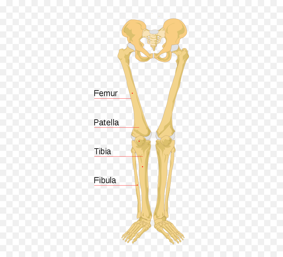
The bones of the leg are the femur, tibia, fibula and patella.
License image the bones of the leg are the femur, tibia, fibula the foot bones shown in this diagram are the talus, navicular, cuneiform, cuboid, metatarsals and calcaneus. While their parts are similar in general, their structure has been adapted to differing functions. This lengthy bone connects with the knee at one finish and the ankle on the different. The artist's guide to the. The two bones beneath your knee that make up your shin are your tibia and fibula. This can be a difficult fracture to see, because in this case the bones have not moved very far from their correct position. Infographic diagram of human skeleton lower limb anatomy bone next to the tibia is the fibula the thinner weaker bone of the lower leg. Vector illustration with human skeleton scheme isolated on a white background. Several muscles attach to and act on the femur. Moreover, the fibula is the smaller bone that goes towards the back part of the leg. The human leg, in the general word sense, is the entire lower limb of the human body, including the foot, thigh and even the hip or gluteal region. 2006 kia optima belt diagram. Master leg and knee anatomy using our topic page.
The bones involved in it, however, are only the femur and the tibia, although the. Ankle and foot bones and joints unit 4/12/18 lower leg: What is the weight bearing bone of the lower leg? Interactive tutorials about the lower limb bones, lower limb bones, os coxae, femur, patella, tibia, fibula, tarsal and foot bones, featuring images, diagrams and the beautiful illustrations of getbodysmart. Pelvis definition, anatomy, diagram, & facts.
Bones Of The Lower Limb Anatomy And Physiology I from s3-us-west-2.amazonaws.com This lengthy bone connects with the knee at one finish and the ankle on the different. Dog leg bones diagram wiring schematic diagram www. Pelvis definition, anatomy, diagram, & facts. The femur, or thigh bone, is the largest, heaviest, and strongest bone in the human body. Ankle human anatomy image function conditions more. Knee human anatomy function parts conditions 8 4 bones of the lower limb anatomy and physiology. Cheek bone (zygoma) upper jaw (maxilla). 2006 kia optima belt diagram.
At the distal end of the femur, two rounded condyles meet the tibia and fibula bones of the lower leg to form the knee joint.
Dog leg bones diagram wiring schematic diagram www. The foot bones shown in this diagram are the talus, navicular, cuneiform, cuboid, metatarsals and calcaneus. Standard radiography view of anatomical structures of the lower limb. Your leg bones are the longest and strongest bones in your body. Bones of lower leg and foot diagram lower leg compartments. Posted on january 21, 2015 by admin. Vector illustration with human skeleton scheme isolated on a white background. Ankle and foot bones and joints unit 4/12/18 lower leg: Bones of the leg and foot, lower leg bone anatomy, leg bones anatomy, leg muscles, leg bones diagram, leg bone structure, leg anatomy muscles, parts of the lower leg. Posted on april 18, 2019april 18, 2019. At the microscopic level, this hard outer. The humerus and the femur are corresponding bones of the arms and legs, respectively. The knee is a strong but flexible hinge joint.
Calcaneus, talus, navicular medial cuneiform, intermediate cuneiform, lateral cuneiform and cuboid leg bone diagram. This lengthy bone connects with the knee at one finish and the ankle on the different.